04. Acute Coronary Syndromes and Regional Myocardial Infarction
Acute transmural myocardial ischemia generates an injury current that produces epicardial ST-segment elevation, reciprocal ST depression, and evolutionary Q waves reflecting the regional anatomical coronary territory.
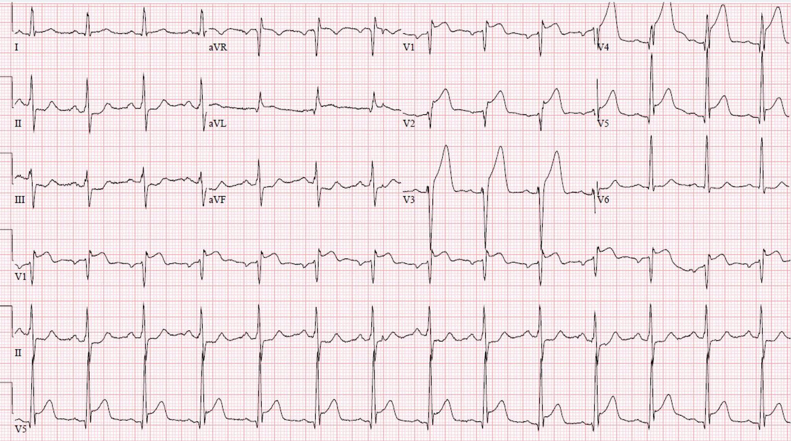 Figure 4.1: Clinical 12-lead ECG of Acute Anterior STEMI displaying marked ST-segment elevation in leads V1-V4 with reciprocal ST depression in inferior leads. Rendered with solid white background.
1. Regional Anatomical Localization of Myocardial Infarction
| Infarct Territory | Primary Leads with ST Elevation | Reciprocal Leads with ST Depression | Culprit Coronary Artery | Critical Clinical Complications |
|---|---|---|---|---|
| Anteroseptal / Extensive Anterior | $V_1 - V_4$ (extensive: I, aVL, $V_1 - V_6$). | II, III, aVF. | Left Anterior Descending (LAD). | Acute cardiogenic shock, ventricular septal rupture, apical aneurysm, LV mural thrombus. |
| High Lateral | I, aVL (occasionally $V_5 - V_6$). | II, III, aVF. | First Diagonal ($D_1$) of LAD or Obtuse Marginal ($OM_1$) of LCx. | Ventricular tachycardia, progressive heart failure. |
| Inferior | II, III, aVF. | I, aVL. | Right Coronary Artery (RCA) ($85\%$) or Left Circumflex (LCx) ($15\%$). | Sinus bradycardia, Mobitz I / Complete AV Block, RV infarction. |
| Right Ventricular (RV) | $V_3R, V_4R$ (STE $\ge 0.5-1.0\text{ mm}$). | Lateral leads. | Proximal Right Coronary Artery. | Severe hypotension with clear lung fields; absolute contraindication to Nitrates. |
| True Posterior | $V_7, V_8, V_9$ (STE $\ge 0.5\text{ mm}$). | $V_1 - V_3$ (Horizontal ST depression, tall wide R, upright T). | Posterior Descending Artery (from RCA or LCx). | Acute mitral regurgitation from posteromedial papillary muscle rupture. |
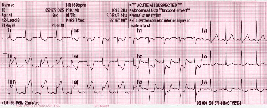 Figure 4.2: 12-lead ECG of Acute Inferior Myocardial Infarction demonstrating ST elevation in leads II, III, aVF with reciprocal depression in leads I and aVL.
2. Differentiating RCA vs LCx Culprit in Inferior STEMI
- RCA Culprit: ST elevation in Lead $\text{III} > \text{Lead II}$ PLUS ST depression in Lead I and aVL $>1.0\text{ mm}$.
- LCx Culprit: ST elevation in Lead $\text{II} \ge \text{Lead III}$ PLUS isoelectric or elevated ST segment in Lead I and aVL.
3. Multimodality Diagnostic Investigations
- High-Sensitivity Cardiac Troponin (hs-cTnI / hs-cTnT): $0/1\text{-hour}$ or $0/2\text{-hour}$ rapid rule-in / rule-out protocols.
- Emergency Transthoracic Echocardiography (TTE): Immediate bedside detection of regional wall motion abnormalities (hypokinesia/akinesia), mechanical complications (free wall rupture, VSR, acute MR).
- Emergency Coronary Angiography (Invasive): Gold standard for culprit lesion identification and mechanical revascularization.
4. Evidence-Based Emergency Management Protocols
- Primary Percutaneous Coronary Intervention (PPCI): Door-to-Balloon time $<90\text{ minutes}$ (or $<120\text{ minutes}$ from first medical contact).
- Dual Antiplatelet Therapy (DAPT):
- Aspirin: $162-325\text{ mg}$ loading dose orally.
- Potent P2Y12 Inhibitor: Ticagrelor ($180\text{ mg}$ load $\to 90\text{ mg}$ bid) or Prasugrel ($60\text{ mg}$ load $\to 10\text{ mg}$ daily). Clopidogrel ($600\text{ mg}$ load) if others contraindicated.
- Anticoagulation: IV Unfractionated Heparin ($70-100\text{ U/kg}$ bolus targeting ACT $250-300\text{ s}$) or Bivalirudin.
- RV Infarction Hemodynamic Protocol: IV crystalloid volume loading ($1-2\text{ L}$); strictly avoid Nitrates, Morphine, and Diuretics.