05. High-Risk STEMI Equivalents and Occlusion Myocardial Infarction
Occlusion Myocardial Infarction (OMI) encompasses acute coronary artery occlusions that do not meet millimeter-based STEMI criteria on standard guidelines, but represent critical transmural myocardial infarction requiring immediate emergent catheterization laboratory activation.
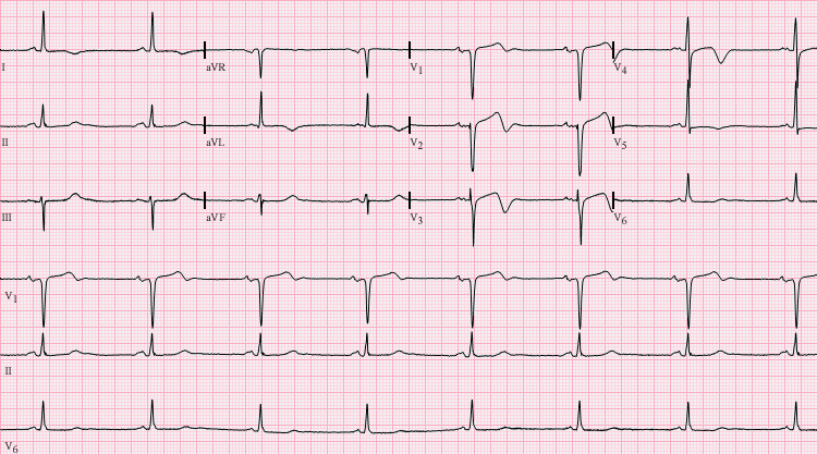 Figure 5.1: Wellens' Syndrome Type B pattern demonstrating deep, symmetrical, inverted T waves across precordial leads V2-V4 during a pain-free interval. Rendered with solid white background.
1. High-Risk STEMI Equivalents Matrix
| STEMI Equivalent Pattern | Characteristic Electrocardiographic Signature | Culprit Coronary Vessel & Pathology | Critical Clinical Management Mandate |
|---|---|---|---|
| Wellens Syndrome | Type A (25%): Biphasic (+/-) T waves in $V_2 - V_3$. Type B (75%): Deep, symmetrical, inverted T waves in $V_2 - V_3$ ($V_1-V_5$). Normal ST segment; zero Q waves. | Critical high-grade proximal LAD stenosis ($>90\%$). Recorded during pain-free interval. | Urgent Coronary Angiography. Stress testing is strictly contraindicated (triggers fatal anterior MI). |
| de Winter T-Wave Pattern | $1-3\text{ mm}$ upsloping ST depression at the J-point continuing into tall, prominent, symmetrical, hyperacute T waves in leads $V_1 - V_6$. | Acute total proximal LAD occlusion without ST elevation. | Immediate Emergent Cath Lab Activation (identical to STEMI). |
| Left Main / 3-Vessel Ischemia | ST-segment elevation in lead aVR $\ge 1\text{ mm}$ ($aVR \ge V_1$) associated with multilead diffuse ST depression ($\ge 1\text{ mm}$) in $\ge 6-8$ leads. | Left Main Coronary Artery (LMCA) total/subtotal occlusion or severe 3-vessel CAD. | Emergent Cath Lab activation; prepare for mechanical circulatory support (Impella/IABP) and CABG. |
| Aslanger Pattern | ST elevation in Lead III only, with ST depression in Lead $V_2$, and isoelectric/slightly elevated Lead II. | Acute inferior OMI in the presence of multi-vessel CAD. | Immediate catheterization. |
| Shark Fin Pattern | Giant R wave merging directly with massive ST elevation creating a single wide triangular wave. | Massive transmural infarction with extensive myocardial involvement. | High risk of acute VF and cardiogenic shock. |
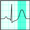 Figure 5.2: de Winter T-wave pattern displaying J-point depression with tall symmetrical T waves in precordial leads.
2. Sgarbossa and Modified Smith-Sgarbossa Criteria in LBBB & Paced Rhythms
Diagnosing acute myocardial infarction in the presence of Left Bundle Branch Block (LBBB) or Right Ventricular Pacing:
MODIFIED SMITH-SGARBOSSA CRITERIA
- Concordant ST Elevation >= 1 mm in any lead with positive QRS (Score: 5)
- Concordant ST Depression >= 1 mm in leads V1, V2, or V3 (Score: 3)
- Excessively Discordant ST Elevation: ST / S Ratio <= -0.25 (ST elevation >= 25% of the preceding S-wave depth)
- Smith Modification: Replaces the unweighted, insensitive absolute $5\text{ mm}$ rule with the proportional $ST/S \le -0.25$ rule, increasing diagnostic sensitivity from $52\%$ to $>91\%$ while maintaining $98\%$ specificity.