01. Fundamental Electrophysiology, Standardization, and Lead Placement
The pediatric electrocardiogram requires rigorous attention to physical calibration, lead geometry, and frequency filtering. Because pediatric voltages can be exceedingly high and heart rates rapid, errors in standardization or electrode placement frequently mimic chamber enlargement or acute conduction failure.
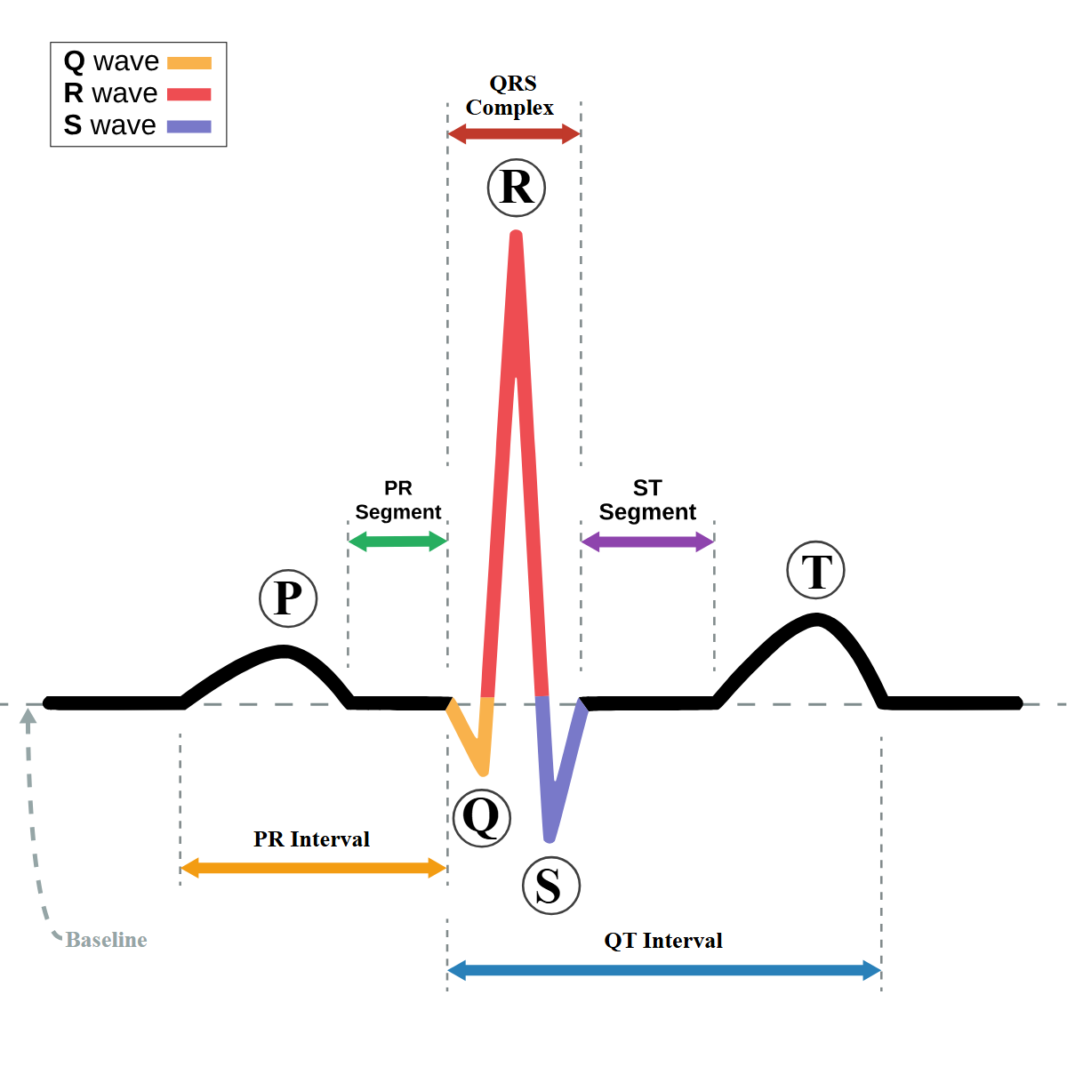 Figure 1.1: Standard surface ECG waveform detailing the P wave, PR interval, QRS complex, ST segment, T wave, and QT interval. Rendered with opaque white background.
1. Clinical Presentation & Bedside Lead Placement
Technical Challenges in Pediatric Lead Placement
- Narrow Intercostal Spaces: In neonates and small infants, standard adult electrodes cause electrical bridging (skin contact overlap). Miniature pediatric suction or silver/silver-chloride disposable electrodes must be used.
- Right-Sided Precordial Leads ($V_3R, V_4R$): Routinely recorded in infants and children presenting with suspected Congenital Heart Disease (CHD), right ventricular pressure/volume overload, or dextrocardia.
- $V_4R$: 5th intercostal space in the right midclavicular line (most sensitive lead for RV hypertrophy and RV ischemia in children).
- $V_3R$: Midway between $V_1$ and $V_4R$.
- Posterior Leads ($V_7 - V_9$): Placed in the 5th intercostal space at posterior axillary, midscapular, and paravertebral lines for complex post-operative anatomy and suspected posterior ventricular ischemia.
Bedside Recognition of Common Lead Misplacements
- Limb Lead Reversal (Right Arm / Left Arm Reversal):
- Inverted P wave, negative QRS complex, and inverted T wave in Lead I.
- Lead aVR exhibits a positive P wave and positive QRS (mimics dextrocardia, but precordial R-wave progression remains entirely normal).
- True Dextrocardia:
- Inverted P and QRS in Lead I PLUS reverse precordial progression (tallest R in $V_1$, fading to flat $rS/QS$ in $V_6$).
- Confirmed by obtaining right-sided precordial leads ($V_1R - V_6R$) which show normal progression.
2. Electrophysiological Principles & Paper Standardization
| Parameter | Standard Speed / Voltage | Double Speed ($50\text{ mm/s}$) | Half Voltage ($5\text{ mm/mV}$) |
|---|---|---|---|
| Paper Speed | $25\text{ mm/s}$ ($1\text{ mm} = 0.04\text{ s}$) | Used in severe neonatal tachycardias ($>250\text{ bpm}$) to unmask hidden P waves. ($1\text{ mm} = 0.02\text{ s}$). | Doubles interval measurements. |
| Voltage Calibration | $10\text{ mm} = 1.0\text{ mV}$ ($1\text{ mm} = 0.1\text{ mV}$) | Used in low voltage tracings (Myocarditis, Pericardial effusion). | Used in extreme ventricular hypertrophy to prevent QRS overlap across lead tracks. |
3. Mathematical Heart Rate Calculation in Children
- Standard 300 / 1500 Rule (Regular Rhythms): $$\text{HR (bpm)} = \frac{300}{\text{Number of large squares between consecutive R-R}} = \frac{1500}{\text{Number of small squares between consecutive R-R}}$$
- Six-Second Strip Method (Irregular Rhythms & Extreme Sinus Arrhythmia): $$\text{HR (bpm)} = (\text{Number of QRS complexes in } 30 \text{ large squares [6.0 s]}) \times 10$$
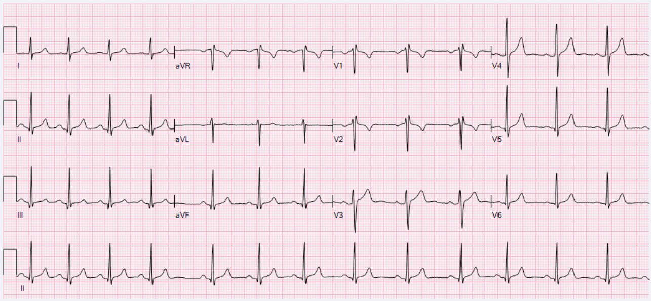 Figure 1.2: Physiological Respiratory Sinus Arrhythmia in a healthy 6-year-old child. P-P and R-R intervals shorten during inspiration and lengthen during expiration.
4. Multimodality Diagnostic Investigations
- Chest Radiography (CXR): Essential when abnormal P/QRS polarities suggest cardiac malposition (situs solitus vs inversus vs ambiguus, levocardia vs dextrocardia vs mesocardia).
- Transthoracic Echocardiography (TTE): Immediate bedside anatomical confirmation of cardiac chambers, systemic/pulmonary venous connections, and great arterial relationships.
5. Evidence-Based Management Protocols
- Artifact Troubleshooting:
- Somatic Tremor / Shivering: Apply low-pass muscle filter ($35-40\text{ Hz}$), ensure child is warm and calm.
- Baseline Wander: Ensure adequate skin preparation, dry skin, replace loose gel electrodes, apply high-pass filter ($0.05-0.5\text{ Hz}$).
- Half-Standardization Interpretation:
- Always multiply recorded amplitudes by $2$ when interpreting voltage criteria on a $5\text{ mm/mV}$ tracing.
6. Clinical Pearls & Diagnostic Traps
Diagnostic Pearl: Before diagnosing sinus pause or AV block in a crying infant, verify lead attachment. A wandering baseline or loose limb lead during active infant movement frequently mimics pauses or polymorphic ventricular tachycardia.