03. Cardiac Axis, Vectorcardiography, and Fascicular Conduction
Frontal plane QRS axis determination is a primary diagnostic gateway in pediatric electrocardiography. In children, axis abnormalities often point directly to specific anatomical congenital defects.
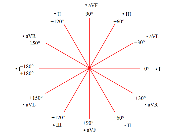 Figure 3.1: The Hexaxial Reference System (Cabrera Circle) displaying limb lead vectors for frontal plane axis determination. Rendered with solid white background.
1. Frontal QRS Axis Determination
The frontal axis is determined using Leads I and aVF:
| Lead I Polarity | Lead aVF Polarity | Frontal QRS Axis | Age-Specific Clinical Significance | Major Differential Diagnoses |
|---|---|---|---|---|
| Positive (+) | Positive (+) | Normal ($0^\circ \text{ to } +90^\circ$) | Normal in children $>1$ year. | Normal heart. |
| Negative (-) | Positive (+) | Right Axis ($+90^\circ \text{ to } +180^\circ$) | Normal in neonates; abnormal in older children. | RVH, Secundum ASD, Tetralogy of Fallot, Cor Pulmonale. |
| Positive (+) | Negative (-) | Left Superior Axis ($-30^\circ \text{ to } -90^\circ$) | Always abnormal at any pediatric age! | Tricuspid Atresia, Primum ASD / AV Canal Defect, Left Anterior Fascicular Block. |
| Negative (-) | Negative (-) | Extreme Northwest ($+180^\circ \text{ to } -90^\circ$) | Always abnormal! | d-TGA with VSD, Severe TOF, Truncus Arteriosus, Ventricular Tachycardia. |
2. Pediatric Fascicular Conduction Blocks
Left Bundle Branch
/
Left Anterior Left Posterior
Fascicle (LAFB) Fascicle (LPFB)
- Axis: -45° to -90° - Axis: > +120°
- qR in I, aVL - rS in I, aVL
- rS in II, III, aVF - qR in II, III, aVF
- Left Anterior Fascicular Block (LAFB): Due to abnormal conduction along the anterior division of the left bundle. In children, it is characteristically seen in Endocardial Cushion Defects (Atrioventricular Septal Defects) due to hypoplasia and posterior displacement of the left anterior fascicle.
- Left Posterior Fascicular Block (LPFB): Rare in isolation in children; requires exclusion of right ventricular hypertrophy.
3. Multimodality Investigations
- Transthoracic Echocardiography (TTE): Mandatory when a left superior axis is detected in an infant to assess for complete atrioventricular canal defect (primum ASD, inlet VSD, common AV valve) or tricuspid atresia with hypoplastic RV.
- Vectorcardiography: Demonstrates a counterclockwise superior frontal loop in AVSD, contrasting with the clockwise loop seen in standard RVH.
4. Evidence-Based Clinical Management
- Surgical Timing for AVSD with Superior Axis: Elective surgical correction is typically performed at $3-6$ months of age before irreversible pulmonary vascular obstructive disease develops.
- Monitoring for Post-Repair Heart Block: Children with AVSD have a displaced, postero-inferior AV node, carrying a lifelong risk of conduction block following surgical patch repair.
5. Clinical Pearls & Diagnostic Traps
Diagnostic Pearl: If a 3-day-old cyanotic neonate presents with a "normal adult axis" ($+30^\circ$) and LV forces, this is profoundly abnormal for a neonate and indicates absent RV mass $\to$ investigate immediately for Tricuspid Atresia or Pulmonary Atresia with Intact Ventricular Septum.