05. Sinoatrial and Atrioventricular Conduction Blocks
Atrioventricular (AV) and sinoatrial conduction delays in pediatric patients range from benign, vagally-mediated nocturnal findings to autoimmune congenital complete heart block requiring urgent pacing.
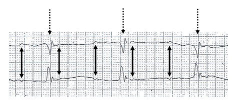 Figure 5.1: 12-lead ECG of Complete Third-Degree AV Block in an infant displaying complete dissociation between P waves and slow, regular QRS complexes. Rendered with solid white background.
1. Clinical Presentation & Bedside Evaluation
- Congenital Complete Heart Block (CCHB): In utero presentation with fetal bradycardia ($HR < 100\text{ bpm}$) or non-immune hydrops fetalis. Postnatally: poor feeding, lethargy, pallor, cannon 'a' waves in the jugular venous pulse, and variable intensity first heart sound ($S_1$).
- Acquired Heart Block: History of tick bite (Lyme carditis), fever/chorea (Rheumatic carditis), recent cardiac surgery (VSD/AV canal patch repair), or myocarditis.
2. Electrocardiographic Classification & Criteria
| AV Block Classification | PR Interval Behavior | Conduction Ratio | Anatomical Site of Block | Clinical Significance |
|---|---|---|---|---|
| First-Degree AV Block | Constant prolongation $>98\text{th}$ percentile for age and rate (e.g. $>0.16\text{ s}$ in young child, $>0.18\text{ s}$ in adolescent). | $1:1$ | AV Node | Minor Jones criterion in Acute Rheumatic Fever; Lyme carditis; Endocardial cushion defects. |
| Second-Degree Mobitz I (Wenckebach) | Progressive PR lengthening until a P wave is non-conducted (dropped QRS). Grouped beating. | Variable ($3:2, 4:3$) | AV Node | High vagal tone in athletic children, nocturnal; usually benign. |
| Second-Degree Mobitz II | Constant PR interval with intermittent, unheralded non-conducted P waves. | Variable ($2:1, 3:1$) | Infranodal (His-Purkinje) | Pathological; high risk of progression to Complete Heart Block. |
| Third-Degree Complete AV Block | Complete AV dissociation; P-P intervals regular (atrial rate normal) and R-R intervals regular (ventricular escape rate slow); PR intervals completely variable. | $0:1$ (No conduction) | AV Node / Infranodal | Congenital autoimmune (Anti-Ro/La) or surgical/infective injury. |
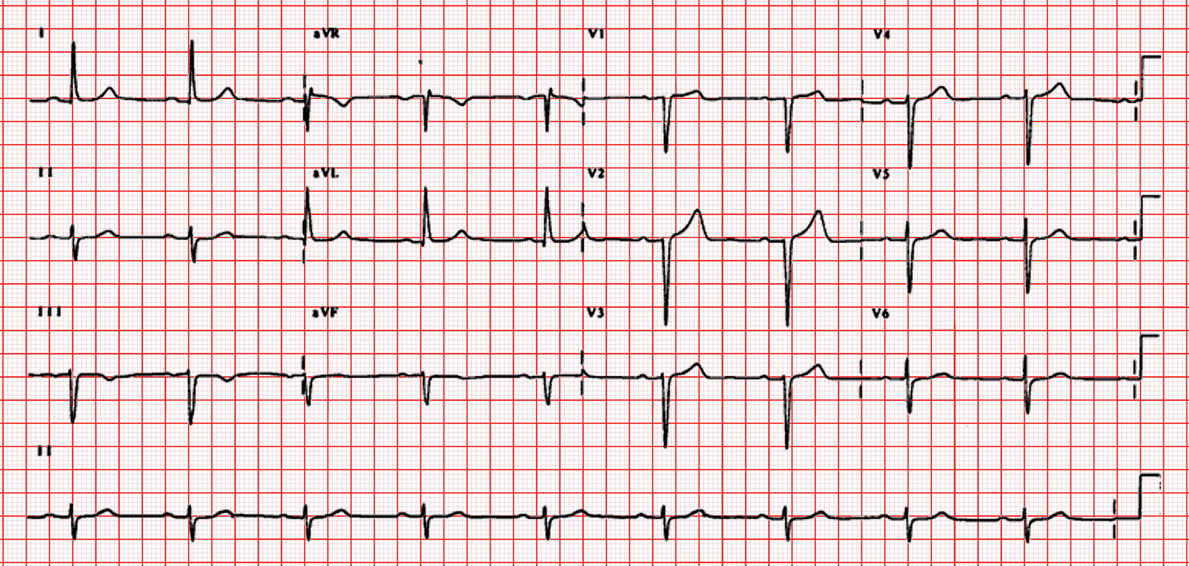 Figure 5.2: First-degree AV block demonstrating constant, prolonged PR interval across all leads.
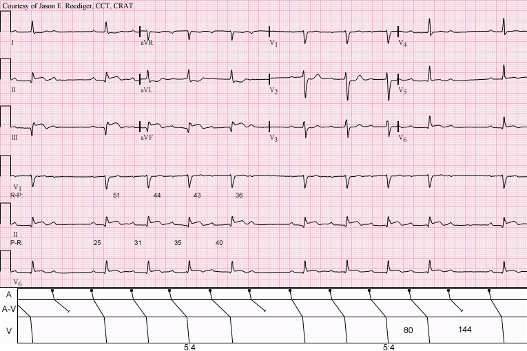 Figure 5.3: Mobitz Type I (Wenckebach) showing progressive PR prolongation culminating in a dropped QRS.
3. Pathophysiology of Congenital Complete Heart Block (CCHB)
- Autoimmune Transplacental Transmission: Maternal autoantibodies (anti-Ro/SSA and anti-La/SSB) cross the placenta between $16-24$ weeks gestation, binding to fetal L-type calcium channels and triggering immune-mediated myocarditis, calcification, and irreversible fibrous replacement of the fetal AV node.
- Structural CCHB: Corrected Transposition of the Great Arteries (L-TGA) and Left Isomerism (Polysplenia syndrome).
4. Multimodality Diagnostic Investigations
- Fetal Echocardiography: Measures mechanical PR interval using pulsed-wave Doppler (mitral inflow / aortic outflow sampling).
- Maternal Serologies: Anti-Ro/SSA and Anti-La/SSB antibody titers.
- 24-Hour Ambulatory Holter Monitoring: Quantifies mean hourly heart rate, pauses, and ventricular escape ectopy.
5. Evidence-Based Management Protocols
- In Utero Therapy (Second-Degree / Early Third-Degree Block): Maternal Dexamethasone ($4\text{ mg/day}$ orally, crosses placenta) $\pm$ IVIG to arrest inflammatory progression.
- Emergency Neonatal Management:
- IV Isoproterenol infusion ($0.05-0.5\ \mu\text{g/kg/min}$) or Epinephrine for symptomatic bradycardia.
- Temporary transcutaneous or transvenous pacing.
- Indications for Permanent Epicardial/Endocardial Pacemaker Implantation in CCHB:
- Symptomatic bradycardia, syncope, or congestive heart failure.
- Infant resting ventricular rate $<50-55\text{ bpm}$ ($<70\text{ bpm}$ with structural CHD).
- Ventricular pauses $>3\times$ basic cycle length.
- Wide QRS escape rhythm, complex ventricular ectopy, or prolonged $QTc$.
6. Clinical Pearls & Diagnostic Traps
Clinical Pearl: Maternal asymptomatic status does not exclude anti-Ro antibodies. More than $80\%$ of mothers whose infants are born with CCHB are completely asymptomatic at the time of delivery.