06. Intraventricular Conduction Delays and Bundle Branch Blocks
Intraventricular conduction delays in pediatric patients reflect disruption within the specialized His-Purkinje network, either congenital, post-surgical, or secondary to ventricular dilatation and strain.
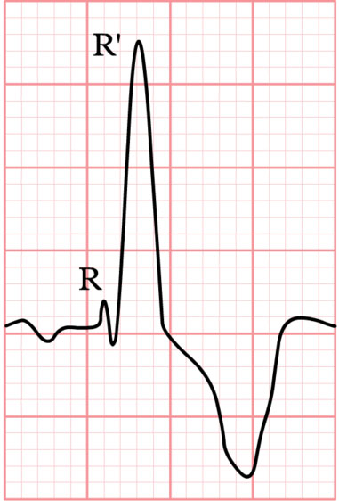 Figure 6.1: Classic Right Bundle Branch Block (RBBB) showing prominent rsR' in V1 and broad slurred S in leads I and V6. Rendered with solid white background.
1. Clinical Presentation & Physical Examination
- Auscultation in RBBB: Wide, fixed splitting of the second heart sound ($S_2$) without respiratory variation in Secundum ASD; wide splitting with normal respiratory variation in isolated RBBB.
- Post-Cardiac Surgical Context: Development of new RBBB following ventricular septal defect patch closure or right ventriculotomy during repair of Tetralogy of Fallot.
2. Diagnostic Electrocardiographic Criteria
| Conduction Disturbance | Precordial Leads ($V_1 - V_2$) | Lateral Leads (I, aVL, $V_5 - V_6$) | QRS Duration Criteria | Common Pediatric Associations |
|---|---|---|---|---|
| Complete RBBB | $rsR'$, $rSR'$, or wide notched $R$ with secondary ST depression and T inversion. | Wide, slurred $S$ wave ($S$ duration $> R$ duration). | $>98\text{th}$ percentile for age ($>0.09-0.10\text{ s}$). | Post-TOF repair, post-VSD repair, Ebstein anomaly, severe RV strain. |
| Incomplete RBBB (IRBBB) | $rsR'$ or $rSr'$ pattern in $V_1$. | Normal terminal S wave. | Normal QRS duration for age ($<98\text{th}$ percentile). | Secundum ASD (RV volume overload), normal variant in healthy infants. |
| Complete LBBB | Broad, deep $QS$ or $rS$ complex with ST elevation. | Broad, notched monophasic $R$ wave without septal $q$ waves; secondary ST depression and T inversion. | $>98\text{th}$ percentile for age. | Dilated Cardiomyopathy, Severe Aortic Stenosis, post-aortic valve surgery. |
| Non-Specific IVCD | Widened QRS complex not meeting classic RBBB or LBBB morphology. | Diffuse widening and notchings. | $>98\text{th}$ percentile for age. | Severe hyperkalemia, severe metabolic acidosis, sodium channel blocker toxicity. |
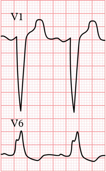 Figure 6.2: 12-lead LBBB showing monophasic notched R waves in lateral leads and QS pattern in lead V1.
3. Multimodality Investigations
- Echocardiography: Assesses ventricular mechanical dyssynchrony, septal flash, and right/left ventricular ejection fraction.
- Cardiac Magnetic Resonance (CMR): In post-TOF patients with RBBB, CMR evaluates right ventricular end-diastolic volume index (RVEDVi) and pulmonary regurgitant fraction.
4. Evidence-Based Management Protocols
- Isolated Asymptomatic RBBB/IRBBB: No specific treatment required; monitor annually.
- Post-Repair Fallot Conduction Monitoring:
- A QRS duration $\ge 180\text{ ms}$ in a post-repair TOF patient is a powerful predictor of malignant ventricular arrhythmias and sudden cardiac death $\to$ triggers evaluation for Pulmonary Valve Replacement (PVR) and ICD therapy.
- Cardiac Resynchronization Therapy (CRT) in pediatric dilated cardiomyopathy with LBBB-induced dyssynchrony and heart failure.
5. Clinical Pearls & Diagnostic Traps
Diagnostic Trap: An $rsR'$ pattern in $V_1$ with normal QRS duration is an incomplete RBBB and does not imply structural conduction injury; it most frequently reflects physiological right ventricular outflow tract conduction delay or volume overload from an uncomplicated Secundum ASD.