07. Ventricular Pre-Excitation and Accessory Pathway Syndromes
Ventricular pre-excitation occurs when an anomalous muscular accessory pathway (bundle of Kent) connects the atrial and ventricular myocardium, bypassing the physiological conduction delay of the AV node.
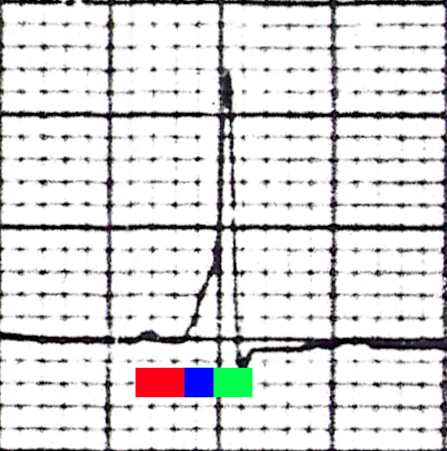 Figure 7.1: Pre-excitation in lead V2 showing the classical triad: short PR interval (<0.10s), slurred upstroke (delta wave), and widened QRS complex. Rendered with solid white background.
1. Clinical Presentation & Bedside Evaluation
- Infants: Present with sudden irritability, pallor, tachypnea, diaphoresis with feeding, and poor perfusion due to paroxysmal supraventricular tachycardia ($HR \approx 250-300\text{ bpm}$).
- Older Children & Adolescents: Sudden-onset paroxysmal palpitations, presyncope, or syncope during physical activity.
- Asymptomatic WPW Pattern: Pre-excitation discovered incidentally on routine ECG screening.
2. Diagnostic Electrocardiographic Hallmarks
- Short PR Interval: Below age-specific lower limit of normal ($<0.08\text{ s}$ in infants, $<0.10\text{ s}$ in young children, $<0.12\text{ s}$ in adolescents).
- Delta Wave: Initial slurring of the QRS complex representing early, slow ventricular myocardial activation via the accessory pathway.
- QRS Prolongation: Widened total QRS duration with secondary ST-T wave changes opposite in direction to the delta wave.
3. Structural Cardiac & Syndromic Associations
| Associated Condition | Clinical & Electrocardiographic Features | Accessory Pathway Characteristics |
|---|---|---|
| Ebstein Anomaly | Massive right atrium, downward displacement of tricuspid leaflets, Himalayan P waves. | Multiple accessory pathways (typically right posterior/posteroseptal); Type B WPW (negative delta in $V_1$). |
| Hypertrophic Cardiomyopathy (HCM) | Massive LV hypertrophy, familial sudden death risk. | Fasciculoventricular connections, Danon disease ($LAMP2$), $PRKAG2$ mutation. |
| Corrected Transposition (L-TGA) | Ventricular inversion, AV block risk. | Left-sided (morphological tricuspid) accessory pathways. |
| Pompe Disease (GSD II) | Severe infantile hypotonia, cardiomegaly. | Extreme high-voltage LVH with extremely short PR interval (pseudo-preexcitation). |
4. Multimodality Diagnostic Investigations
- Holter Monitoring: Documents intermittent pre-excitation (which suggests a long refractory period and lower sudden death risk).
- Exercise Treadmill Test (ETT): Sudden, abrupt loss of the delta wave at higher heart rates indicates a long accessory pathway refractory period (low-risk pathway).
- Invasive Electrophysiology Study (EPS): Determines the Shortest Pre-Excited R-R Interval (SPERRI) in Atrial Fibrillation. A $\text{SPERRI} < 250\text{ ms}$ defines a high-risk accessory pathway capable of precipitating ventricular fibrillation.
5. Evidence-Based Clinical Management Protocols
- Acute Orthodromic Tachycardia:
- Vagal maneuvers (ice bag to the face in infants for $10-15\text{ s}$, modified Valsalva in older children).
- IV Adenosine: Rapid push $0.1\text{ mg/kg}$ (max $6\text{ mg}$), increase to $0.2\text{ mg/kg}$ (max $12\text{ mg}$).
- Synchronized DC Cardioversion ($0.5-1.0\text{ J/kg}$) if hemodynamically compromised.
- Pre-Excited Atrial Fibrillation (Emergency):
- Contraindicated Drugs: AV nodal blockers (Adenosine, Digoxin, Verapamil, Beta-blockers) are strictly contraindicated due to preferential conduction down the accessory pathway leading to VF.
- Definitive Acute Treatment: Synchronized DC Cardioversion or IV Procainamide.
- Definitive Curative Catheter Ablation:
- Radiofrequency (RF) or Cryoablation: Class I indication for symptomatic WPW, high-risk pathway features ($ ext{SPERRI} < 250\text{ ms}$), or structural CHD. Cryoablation is preferred for septal pathways adjacent to the normal AV node to eliminate the risk of permanent complete heart block.
6. Clinical Pearls & Diagnostic Traps
Red Flag Warning: Never administer Digoxin or Verapamil to an infant or child with WPW. Both drugs shorten accessory pathway refractory periods and can trigger fatal ventricular fibrillation.