12. Cyanotic Congenital Heart Disease Signatures
Cyanotic congenital heart lesions involve right-to-left intracardiac or great vessel shunting, causing arterial hypoxemia and unique chamber pressure-volume remodeling patterns.
1. Clinical Presentation & Physical Examination
| Cyanotic Lesion | Cardinal Bedside Signs | Murmurs & Auscultatory Findings |
|---|---|---|
| Tetralogy of Fallot (TOF) | Central cyanosis, clubbing, squatting posture, hypercyanotic ("tet") spells. | Single loud $S_2$, harsh ejection systolic murmur at mid-left sternal border (RVOT obstruction). |
| Transposition of Great Arteries (d-TGA) | Severe central cyanosis within first hours of life, unresponsive to hyperoxia test. | Single loud $S_2$; often no significant murmur unless VSD is present. |
| Tricuspid Atresia | Progressive cyanosis, absent right-sided pulses, prominent jugular 'a' waves. | Single $S_2$, holosystolic murmur of VSD or continuous murmur of PDA. |
| Ebstein Anomaly | Cyanosis, right heart failure, massive hepatomegaly, superficial venous distension. | Multiple heart sounds (triple/quadruple rhythm), wide split $S_1$ ("sail sound"), holosystolic TR murmur. |
| Total Anomalous Pulmonary Venous (TAPVC) | Severe tachypnea, pulmonary edema (obstructed TAPVC), cyanosis. | Quadruple gallop rhythm, pulmonary systolic flow murmur. |
2. Electrocardiographic Signatures Matrix
| Cyanotic Lesion | Frontal QRS Axis | Chamber Enlargement | Characteristic Diagnostic Hallmark |
|---|---|---|---|
| Tetralogy of Fallot | Right ($+120^\circ \text{ to } +180^\circ$) | Pure RVH | Tall monophasic $R$ in $V_1$ with sudden transition to $rS$ in $V_2$ ("sudden transition" sign); absent LV forces. |
| d-TGA | Right ($+90^\circ \text{ to } +180^\circ$) | RVH (or BVH if VSD) | Marked neonatal RVH that fails to regress; upright T in $V_1$ after day 3. |
| Tricuspid Atresia | Left Superior ($-30^\circ \text{ to } -90^\circ$) | Pure LVH + RAE | Cyanotic neonate + Superior Axis + LVH = Tricuspid Atresia until proven otherwise! |
| Ebstein Anomaly | Normal or Right | RAE + RBBB | "Himalayan" P waves ($>5\text{ mm}$), splintered low-voltage RBBB in $V_1$, accessory pathways (WPW). |
| TAPVC | Marked Right ($+150^\circ \text{ to } +180^\circ$) | Severe RVH + RAE | $qR$ pattern in $V_1$ with peaked P-pulmonale; RV strain pattern. |
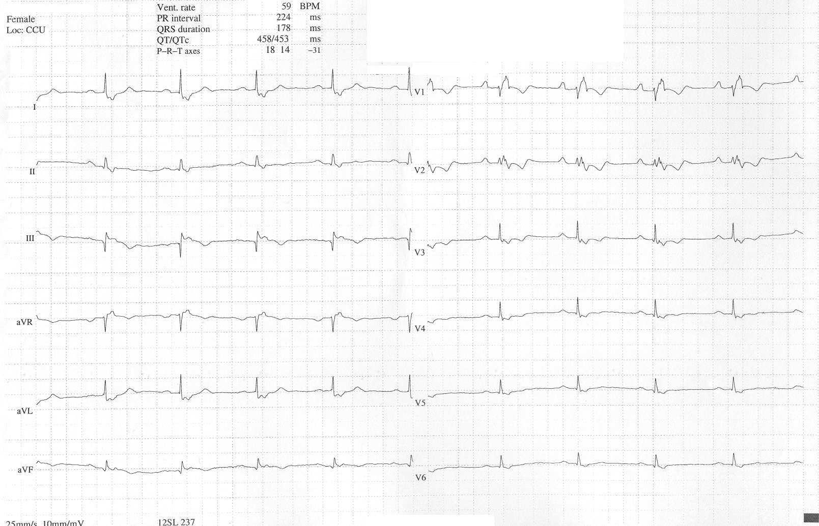 Figure 12.1: Clinical 12-lead ECG in Ebstein Anomaly displaying giant Himalayan P waves in Lead II and V1, low-voltage splintered RBBB in V1, and first-degree AV block. Rendered with solid white background.
3. Multimodality Investigations
- Hyperoxia Test: $PaO_2 < 150\text{ mmHg}$ on $100\% \ O_2$ confirms intracardiac cyanotic shunt.
- Echocardiography (TTE): Defines great arterial spatial alignment (parallel outflows in TGA), ventricular septal anatomy, RVOT obstruction degree, and tricuspid valve displacement index.
- Chest Radiography (CXR): Boot-shaped heart (TOF), Egg-on-a-string (TGA), Snowman / Figure-of-8 (supracardiac TAPVC), Box-shaped massive cardiomegaly (Ebstein).
4. Evidence-Based Emergency & Surgical Management
- Ductal-Dependent Lesions: Immediate IV Prostaglandin E1 ($PGE_1$ / Alprostadil) infusion ($0.01-0.05\ \mu\text{g/kg/min}$) to maintain ductal patency.
- Hypercyanotic ("Tet") Spell Protocol:
- Knee-chest position (increases systemic vascular resistance, reducing right-to-left shunt).
- High-flow oxygen ($100\%$).
- IV Morphine Sulfate ($0.1-0.2\text{ mg/kg}$ SC/IM/IV) to suppress respiratory center hyperpnea.
- IV Sodium Bicarbonate ($1-2\text{ mEq/kg}$) to correct metabolic acidosis.
- IV Phenylephrine ($5-20\ \mu\text{g/kg}$) or Propranolol ($0.1\text{ mg/kg}$ slow IV).
- Corrective Surgery: Arterial Switch Operation (Jatene) for TGA within first $2-3$ weeks of life; Complete primary repair for TOF at $3-6$ months.