18. Electrolyte Derangements and Metabolic Crises
Electrolyte disturbances directly disrupt the resting membrane potential, phase 0 depolarization velocity, and phase 3 repolarization of pediatric cardiomyocytes, creating life-threatening electrical instability.
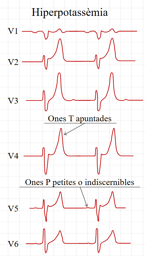 Figure 18.1: Stepwise electrophysiological progression of Hyperkalemia from peaked tented T waves to QRS widening, loss of P waves, and sine wave degeneration. Rendered with solid white background.
1. Potassium Disturbances
| Serum Level | Electrocardiographic Hallmarks | Electrophysiological Mechanism | Emergency Management |
|---|---|---|---|
| Mild Hyperkalemia ($5.5 - 6.5\text{ mEq/L}$) | Tall, narrow-based, symmetrical, peaked/tented T waves in precordial leads. | Accelerated phase 3 repolarization ($I_{Kr}$ activation). | Dietary restriction, loop diuretics, oral potassium binders. |
| Moderate Hyperkalemia ($6.5 - 7.5\text{ mEq/L}$) | PR prolongation, flattening and loss of P wave, widening of QRS complex. | Inactivation of voltage-gated $Na^+$ channels ($I_{Na}$). | Shift $K^+$ intracellularly: IV Regular Insulin ($0.1\text{ U/kg}$) + $25\% \ Dextrose$ ($2\text{ mL/kg}$) + Nebulized Salbutamol. |
| Severe Hyperkalemia ($>8.0\text{ mEq/L}$) | Wide QRS merges with T wave forming a Sine Wave Pattern $\to$ ventricular fibrillation / asystole. | Complete loss of resting membrane excitability (Immediate Resuscitation). | IV Calcium Gluconate 10% ($0.5-1.0\text{ mL/kg}$ over $3-5\text{ min}$) for immediate myocardial membrane stabilization. |
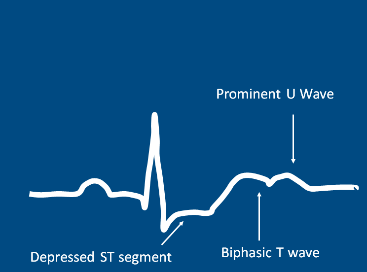 Figure 18.2: Clinical ECG of Hypokalemia displaying ST segment depression, flattened T waves, and prominent pathological U waves in leads V2-V4.
- Hypokalemia ($K^+ < 3.5\text{ mEq/L}$):
- ST segment depression $>0.5\text{ mm}$.
- T-wave flattening or inversion.
- Emergence of prominent U waves ($U$ wave amplitude $> T$ wave in $V_2-V_4$).
- Pseudo-prolongation of QT interval (actually $QU$ interval) predisposing to Torsades de Pointes.
- Treatment: IV Potassium Chloride infusion (max rate $0.5\text{ mEq/kg/hr}$ via peripheral line with continuous cardiac monitoring).
2. Calcium Disturbances
| Derangement | $QTc$ Interval Change | Pathognomonic ECG Pattern | Clinical Context |
|---|---|---|---|
| Hypocalcemia | Prolonged $QTc$ | Isolated lengthening of the isoelectric ST segment with normal T-wave duration and morphology. | Neonatal hypocalcemic tetany, DiGeorge syndrome (22q11 deletion), hypoparathyroidism. |
| Hypercalcemia | Shortened $QTc$ | Abrupt takeoff of T wave directly from the QRS complex with virtually absent ST segment; Osborn J waves. | Williams syndrome (infantile hypercalcemia), hyperparathyroidism, prolonged immobilization. |
3. Multimodality Investigations
- Arterial Blood Gas (ABG): Concomitant metabolic acidosis worsens hyperkalemia by shifting potassium out of cells; severe alkalosis worsens hypokalemia.
- Ionized Calcium & Serum Magnesium: Mandatory in any child with unexplained $QTc$ prolongation.